
Explainer · August 10, 2026 · 4 min · By Marisol Etcheverry
Why a Treated Age Spot Can Come Back Darker, and What That Actually Means
Post-inflammatory hyperpigmentation, true recurrence, and misdiagnosed lesions all look similar in the mirror. The biology behind each one is different, and so is the fix.
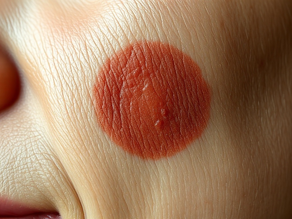
One of the most common frustrations in age spot treatment is the spot that seems to vanish, then returns weeks or months later, sometimes darker than before. Patients often assume the procedure failed or that the pigment is somehow permanent. In most cases neither is true. There are three distinct biological explanations, and telling them apart matters because each calls for a different response.
First, understand what an age spot is. A solar lentigo is not a stain sitting on top of the skin. It is a patch of skin where years of ultraviolet exposure have pushed melanocytes, the pigment-producing cells in the basal layer of the epidermis, into a state of chronically elevated activity. These cells hand off extra melanin to surrounding keratinocytes, which carry the pigment upward as skin turns over. Most treatments, including Q-switched and picosecond lasers, intense pulsed light, cryotherapy, and chemical peels, work by destroying or shedding the pigment-loaded keratinocytes. Critically, many of these treatments do not eliminate the overactive melanocytes underneath. The factory often survives even when the inventory is cleared.
Scenario one: post-inflammatory hyperpigmentation. Any treatment that removes pigment does so by creating a controlled injury, and injury triggers inflammation. Inflammatory signaling molecules, including prostaglandins and various cytokines, directly stimulate melanocytes to produce more melanin. This is post-inflammatory hyperpigmentation, usually shortened to PIH. It typically appears two to four weeks after treatment as a brown or gray-brown patch in the treated area, and it is far more common in medium to deeper skin tones, roughly Fitzpatrick types III to VI. PIH is not a failure of the original treatment. It is a separate, temporary pigment response, and it generally fades over three to twelve months, faster with strict sun protection and topical agents such as azelaic acid, retinoids, or hydroquinone under medical guidance. The main clue is timing: darkening that shows up within the first month, often slightly larger or blurrier than the original spot, is usually PIH.
Scenario two: true recurrence. Because the melanocytes in a lentigo remain hyperactive after most treatments, renewed ultraviolet exposure can restart pigment production in the exact footprint of the original spot. This tends to happen more gradually, over several months to a couple of years, and the recurring spot usually matches the original in size and shape. Recurrence rates after laser and IPL treatment are meaningfully higher in people who do not use daily broad-spectrum sunscreen, and visible light plays a role too, which is why dermatologists increasingly recommend tinted sunscreens containing iron oxides for pigment-prone patients. Recurrence is not a sign the treatment was done poorly. It reflects the underlying biology: the treatment cleared pigment, the sun re-triggered the cells that make it.
Scenario three: it was never a simple age spot. Flat brown lesions on sun-exposed skin are not all lentigines. Seborrheic keratoses, which are benign but structurally different, often respond poorly or unevenly to pigment lasers and can appear to darken or thicken afterward. More importantly, lentigo maligna, an early form of melanoma that occurs on chronically sun-damaged skin, can closely mimic a benign age spot. A pigmented lesion that repeatedly returns after treatment, grows, develops irregular borders, or shows multiple shades of brown and black should be evaluated by a dermatologist, and a biopsy may be appropriate before any further cosmetic treatment. This is one reason clinicians are cautious about treating ambiguous lesions with destructive methods that leave no tissue to examine.
How clinicians reduce the odds of rebound darkening. Several strategies are well supported by the mechanism. Conservative energy settings and test spots reduce inflammatory load, especially in darker skin. Some practitioners pre-treat and post-treat pigment-prone patients with topical agents that suppress melanocyte activity, including hydroquinone, azelaic acid, or tranexamic acid formulations, to blunt the PIH response. Spacing sessions further apart allows inflammation to fully resolve before the next injury. And the single highest-yield intervention is unglamorous: daily broad-spectrum sunscreen, SPF 30 or higher, reapplied during extended sun exposure, ideally tinted for visible light protection, continued indefinitely.
The bottom line. A spot that darkens within the first month after treatment is most often PIH and usually resolves with time and sun protection. A spot that fades fully and then slowly returns months later is usually true recurrence driven by surviving melanocytes and new UV exposure, and it can typically be retreated. A spot that behaves unpredictably, changes shape, or keeps coming back despite good sun habits deserves a diagnostic look before another round of treatment. None of these outcomes means age spots are untreatable. They mean the treatment addressed the pigment while the machinery that produces it remained in place, which is exactly what the underlying biology predicts.
Related reading: Why a Zapped Age Spot Can Come Back Darker: The PIH Problem Explained.
More in Explainer
View all →- What Are Age Spots, Really? Three Definitions, and Where They Disagree
- IPL vs. Pigment Lasers for Age Spots: What Actually Differs Under the Skin
- IPL vs. Q-Switched Lasers for Age Spots: What Actually Separates the Two
- IPL, Q-Switched, or Picosecond: How the Three Main Light Treatments for Age Spots Actually Differ